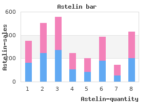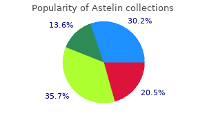
"Discount astelin 10 ml mastercard, allergy symptoms while pregnant". A. Inog, M.B. B.CH., M.B.B.Ch., Ph.D. Co-Director, Creighton University School of Medicine Candida infection can present as isolated white plaques allergy shots injection sites discount astelin 10 ml online, which can be confused with glycogenic acanthosis or progress to form confluent ulcerations with an overlying membrane allergy treatment in dubai purchase astelin 10ml with visa. Herpesvirus tends to produce vesicles or isolated superficial ulcers allergy forecast pa purchase 10ml astelin visa, but extensive involvement can produce confluent ulcerations allergy shots needle size astelin 10ml online. Biopsy of the ulcerated area usually shows either invasive hyphae of Candida or characteristic nuclear changes of the squamous cells when herpesvirus is present. For mild, noninvasive candidal disease, topical therapy with nystatin (250,000 units every 2 hours) or clotrimazole (dissolved in the mouth 5 times per day) suffices. For more serious infections, systemic treatment with oral fluconazole or occasionally ketoconazole is used. Low-dose intravenous amphotericin may be needed for patients who do not respond to oral treatment and those unable to swallow medications. Esophageal Injuries Caustic Ingestion Caustic burns of the esophagus occur in children by accident; adults usually suffer such burns because of suicide attempts. Lye crystals, and especially liquid lye preparations for drain cleaning, detergents, and bleach are the most common causes (see Chapter 98). The speed of lye injury is so great that attempts to neutralize the caustic are futile. The history is all-important, but the degree of esophageal injury must be assessed emergently by endoscopy. Significant esophageal damage has been seen even without oral burns; conversely, oral burns do not necessarily mean that the material has reached the esophagus. If no esophageal reaction is present after apparent caustic ingestion, further care directed toward the esophagus will not be necessary. Circumferential burns and ulcers in the esophagus may result in delayed perforation over several days or in stricture formation. The goals of therapy are to prevent perforation and to avoid progressive fibrosis and stricture of the esophagus. However, the accepted therapy of a definite lye or caustic burn remains unsupported by clinical trials. For burns with solid lye or other solid agents, corticosteroids have been recommended at an initial dose of 80 mg/day, tapering to 20 mg/day until the esophagus heals. For liquid lye, serious consideration should be given to emergent esophagogastrectomy because lesser measures have met with unacceptably high mortality. Damage by Medication Ingested pills may lodge in the esophagus and damage the mucosa in a localized area. Tetracycline, doxycycline, potassium tablets, ascorbic acid, quinidine, and nonsteroidal anti-inflammatory agents are the principal medications that cause pill-induced esophagitis, but the list is long. For example, the bisphosphonates, such as alendronate, also cause esophageal injury. Normal individuals can retain small capsules in the esophagus even when swallowing in the upright position. The clinical syndrome consists of steady burning or chest pain accompanied by local odynophagia occurring 4 to 6 hours after ingesting one of the offending capsules or tablets. Endoscopy usually shows a localized mucosal ulcer, which may heal without scar or lead to a stricture requiring dilation. Pills should be taken by the patient in the upright position, with several swallows of water taken both before and after pill ingestion. Esophageal Trauma the esophagus is well protected by the thoracic cage, but it can be involved either by blunt trauma. Iatrogenic perforation with an endoscope, dilator, or, very rarely, nasogastric tube can lead to similar complications. The mucosal lesion first described by Mallory and Weiss has been recognized much more frequently since the advent of emergency fiberoptic endoscopy. Classically, the patient has repeated attacks of retching, at first producing gastric contents and later bright red blood. One quarter of patients shown to have a Mallory-Weiss tear have no prior history of vomiting. The tear is usually in the gastric mucosa just below the gastroesophageal junction, although it can extend through the junction and up into the esophageal mucosa. The chromosomal disorders should be considered as a possible explanation for multiple anomalies allergy testing eosinophilic esophagitis safe astelin 10 ml, mental retardation allergy shots lincoln ne buy astelin 10ml low cost, recurrent miscarriages allergy testing greenville nc discount astelin 10 ml otc, and unexplained stillbirths (see Chapter 34) allergy natural cure generic astelin 10 ml. Empirical figures must be used to predict the recurrence of chromosomal abnormalities in a family. These range between 1% and 10%, and the literature must be consulted with reference to the specific situation. When a clear mendelian pattern is seen and the disorder is a recognized mendelian condition, counseling is based on that pattern. When the family history fails to demonstrate a mendelian pattern, the diagnosis is reviewed and the medical literature consulted to determine the inheritance pattern for the specific disorder. In autosomal recessive conditions the birth of an affected child may be the first signal that a set of parents is heterozygous for a rare recessive condition. Here the genetic model depends on the correct diagnosis and the known inheritance pattern for that disorder. For X-linked conditions, the decision must be made whether the affected individual represents a new mutation or inheritance from a heterozygous mother who by chance has no affected relatives. In the past, Bayesian calculations based on the pedigree have been the mainstay of this kind of analysis. Today, however, molecular diagnostic tools have refined the ability to determine heterozygosity in this situation. For dominantly inherited conditions, the literature must be consulted to determine the proportion of patients who represent new mutations, a figure that can approach 50%. When a new mutation appears to be the explanation, others in the family are not at risk, but each offspring of the affected individual has a 50% risk of inheriting the gene. Variable expression can confound the analysis of a family demonstrating an autosomal dominant condition. Gonadal mosaicism for the mutation accounts for rare recurrences in families in which neither parent is affected with the dominant condition and no test can exclude it. In general, however, the absence of the condition in any other family member makes the likelihood high that the patient represents a new mutation. Frequently, no mendelian hypothesis can be sustained yet there is familial aggregation of the disorder. Many conditions, such as neural tube defects and cleft lip and palate, appear to be multifactorial in origin with both genetic and environmental components. Genetic counseling for these conditions must rely on empirical figures for the specific condition. The process of genetic counseling itself has the following components: transferring information about the genetic risks, putting the risks in perspective, providing a summary of the disorder, and discussing the options. An explanation of the genetic risks requires imparting factual information using scientific concepts that are not familiar to everyone. It is important that the facts on which the genetic model is based be clearly explained. However, it is neither possible nor desirable to present an entire course in medical genetics to the anxious patient and family. The strategy of first presenting a brief summary of the conclusions and their implications, stating that the evidence for this conclusion will presently be discussed, can allay some fears and relieve some of the distraction that prevents families from hearing this kind of information. If the condition is a chromosome disorder, the structure and ways of identifying chromosomes must be mentioned and the specific disorder illustrated. Using teaching aids such as diagrams and photographs of chromosomes is helpful, with the normal situation providing a frame of reference. When the condition is a mendelian disorder, the basic concepts of single gene inheritance must be discussed briefly, but the discussion should center on the mode of inheritance involved in the particular family and not be clouded with a great deal of extraneous material about other modes of inheritance. Families without a prior family history of the disorder may have difficulty with the fact that the disorder has never been seen in their family. An explanation of heterozygosity may help clarify autosomal recessive inheritance. Autosomal dominant inheritance is easy to understand when there are other affected individuals and the pedigree demonstrates a clear vertical pattern. As in the chromosome disorders, the use of such teaching aids as gene diagrams, sample pedigrees, and other models may be extremely valuable. A second important component of genetic counseling is putting the risk in perspective. This perception depends on at least two factors: (1) risk compared to background risk and (2) overall burden, a combination of risk and severity.
The data can be used to derive a rather precise and predictable log-dose response curve for most agents allergy shots numbness arm buy astelin 10ml. In contrast allergy shots make you sick buy discount astelin 10ml on-line, the toxicologist or epidemiologist studying clinical effects of naturally occurring toxins has none of this information and thus labors under several disadvantages allergy treatment new purchase 10ml astelin with amex. First allergy medicine list in india 10 ml astelin sale, he or she often does not know with certainty the concentration of the toxic agent that was present in the environment when the pathology was induced. Ex-post facto estimations of these exposure concentrations are often a limitation of the science, even if good analytic techniques for the toxin are available. These and other factors necessarily introduce significant uncertainty in the development of dose-response curves for many toxic substances. In view of these limitations, toxicologists who develop permissible levels, "reference doses" (RfDs) for general population exposures to chemicals with known toxic effects, routinely build in large safety factors. The intrinsic difficulties encountered with exposure assessment and outcomes evaluation in the clinical setting may help explain the long controversy and delays involved in validating hypotheses about whether cigarette smoking causes lung cancer, as well as ongoing controversies (such as the putative relationships between electromagnetic fields and cancer and between silver amalgam dental fillings and disease). Proving cause-and-effect relationships for clinical diseases potentially resulting from mycotoxins has additional limitations. There are no standardized methods for qualitative or quantitative analysis of airborne mycotoxins in the indoor (or outdoor) environment, and there are few known biomarkers for measuring exposure to these toxins (Cloeren 2002). Neither of these measurements provides a direct assessment of mycotoxin levels because mycotoxin concentrations may not necessarily correlate with either the total volume of fungal material or the total number of viable spores. Given these limitations, what then can we conclude with respect to mycotoxins and human disease? Toxicity from Ingestion of Mycotoxins the clearest evidence that mycotoxins can cause human disease derives from the effects noted after ingestion of fungus-contaminated food. It was caused by the ingestion of rye or other grain infested with fungi (Claviceps purpura) containing "ergot," which is a complex and variable mixture of alkaloids. Some of these alkaloids are vasoconstrictors, and their ingestion can lead to blistering, gangrene, and loss of limbs in some patients. Consumption of ergot can also result in neuropsychiatric effects, including bizarre behavior, hallucinations, dementia, and convulsions. It has been speculated that such behavioral changes induced by ergot poisoning led to the Salem witch trials in 1692. Outbreaks of ergotism have occurred as recently as 1951, when over 200 persons in Provence, France, developed severe symptoms; 32 went insane, and 4 died from eating bread made from contaminated rye (University of Georgia 2001). Consistent with these clinical effects, there is evidence for neurotoxicity from mycotoxins in sheep and cattle that consumed contaminated feed (Mantle et al. B3 Mycotoxin ingestion also has been implicated in carcinogenesis (Sorenson 1999). Clinically, it has been identified as causing hepatic carcinomas in patients who ingest it in contaminated grain or peanuts, particularly if they have a coexisting hepatitis B infection. These effects include inhibition of phagocytosis, microbiocidal activity, and cytokine production by human monocytes (Cusumano et al. In agreement with these observations, veterinary reports of animals that ingest aflatoxin found in moldy hay have documented suppressed cell-mediated immune responses with reduced phagocytosis and depressed production of complements and interferon. Acquired immunity from vaccination programs has also been shown to be substantially suppressed (Pier 1992). Two episodes of severe aflatoxin poisoning were reported in horses, with encephalomalacia of cerebral hemispheres, fatty degeneration, necrosis, bile duct hyperplasia, fibrosis of the liver, fatty infiltration of the kidney, hemorrhagic enteritis, and myocardial degeneration. The diagnosis was based on gross and histopathologic observations, consistent with observations of other species poisoned with aflatoxin, and on isolation of the toxin from feed and animal tissues (Angsubhakorn et al. It is associated with ingestion of foodstuffs made from barley that was not dried after harvest and was stored through the fall and winter in moist conditions, typically in Yak-skin and Yak-hair bags (Allander 1994, Haubruge et al. This food-related disease has occurred sporadically in Russia, probably since the nineteenth century. Various reports indicate that chronic consumption of grain contaminated with a trichothecene (T-2) mycotoxin resulted in a mortality rate of 10-60 percent of the local population during the years 1942-1947 (Locasto et al. The first phase develops within 72 hours of initial consumption of the contaminated foodstuffs. It results in gastrointestinal inflammation leading to abdominal pain, nausea, and vomiting, often accompanied by headache, weakness, fatigue, and tachycardia.
In poorly controlled diabetes mellitus allergy testing dogs buy astelin 10ml lowest price, the glucosuria and resulting osmotic diuresis increase urinary phosphate loss allergy shots medicare purchase astelin 10 ml on-line. However allergy medicine 8 month old astelin 10ml with mastercard, serum phosphorus is not generally depressed when poorly controlled diabetics are initially evaluated allergy medicine 3 month old purchase astelin 10 ml line, probably because phosphate shifts to the extracellular compartment from the intracellular space. Only after starting therapy with insulin and intravenous fluids does the hypophosphatemia become manifested. In alcoholics, hypophosphatemia and phosphate depletion are caused by multiple factors: the phosphaturic effects of ethanol and magnesium depletion, poor dietary intake, and ketoacidosis. Other contributing factors can be vomiting, diarrhea, and the use of phosphate-binding antacids. This constellation of disorders results in disturbed function of multiple body systems. When total body phosphorus stores are normal, phosphate supplementation is unnecessary (Table 222-3). When body phosphate stores are reduced, urinary losses need to be minimized, gastrointestinal absorption needs to be enhanced, and phosphate supplements may be necessary. To replete body phosphorus stores, 1000 to 2000 mg of phosphorus may need to be supplemented daily for up to 2 weeks. Whenever phosphate replacement is given, serum calcium, magnesium, phosphorus, and electrolytes should be monitored closely. The complications of administering phosphate include diarrhea (after oral administration), hypocalcemia, metastatic calcification, hypotension, hyperkalemia and/or hypernatremia, and metabolic acidosis. Crook M, Swaminathan R: Disorders of plasma phosphate and indications for its measurement. A well-referenced review of the clinical disorders of phosphate and their pathophysiology, manifestations, and treatment. Alfrey Magnesium is the second most common intracellular cation, with only three other cations-potassium, calcium, and sodium-occurring with greater abundance in the body. It plays a crucial role in storing and using energy inasmuch as all enzymatic reactions involving adenosine triphosphate frequently require magnesium. Because magnesium is also an essential element for plants in that it is a constituent of chlorophyll, it is present in virtually all food sources. Despite this wide distribution, the average dietary intake of magnesium is about 25 mEq/day, which only marginally meets the recommended daily requirements for this element. Fractional absorption of magnesium varies from 80% on a magnesium-restricted diet to less than 10% when large oral loads of magnesium are consumed. Therefore, small changes in serum magnesium levels are accompanied by rather rapid increases or decreases in urinary magnesium excretion. The prevalence of hypomagnesemia in a general hospital setting has been estimated to range from 6. Clinical findings of severe hypomagnesemia are mainly confined to the neuromuscular system and consist of muscle fasciculations and tremors, positive Chvostek and Trousseau signs, overt tetany, weakness, anorexia, apathy, and rarely seizures. The biochemical findings of symptomatic hypomagnesemia are serum magnesium levels usually less than 1 mEq/L in association with hypokalemia and hypocalcemia. Magnesium depletion can result from either gastrointestinal or renal causes (Table 223-1). When serum magnesium falls only slightly and if the kidneys respond normally, urinary magnesium excretion falls to less than 12 mg (1 mEq) per day. Therefore, urine magnesium is low if magnesium depletion results from gastrointestinal causes; however, urinary magnesium excretion is in the normal range (120 to 160 mg/day) if depletion results from a renal leak. The fecal magnesium content correlates with the amount of stool fat, which suggests that magnesium malabsorption is a result of magnesium forming an insoluble complex with fat in the gastrointestinal tract. Any severe diarrheal state such as ulcerative colitis, amebic colitis, and intestinal resection can also deplete magnesium. Another gastrointestinal cause of magnesium depletion is an isolated defect in magnesium absorption that usually occurs in infants. Magnesium depletion can rarely result from poor intake as found in protein-calorie malnutrition and patients receiving total parenteral nutrition without magnesium supplementation. Renal magnesium wasting (see Table 223-1) can result from an intrinsic disorder of the renal tubule or from extrinsic or reversible factors. Drugs that most commonly cause magnesium depletion are aminoglycosides, cyclosporine, pentamidine, foscarnet, and cis-diamminedichloroplatinum (cisplatin). Conversely allergy treatment coughing discount astelin 10 ml with amex, chest pain can result from a variety of causes of myocardial ischemia other than atherosclerosis or from non-ischemic causes of cardiac or non-cardiac origin allergy cold purchase 10ml astelin with visa. Angina may be defined by the stability or non-stability of its manifestation allergy shots length of treatment purchase astelin 10 ml with mastercard, its provocation factors allergy shots memphis tn purchase astelin 10ml with mastercard, or its pathophysiology. Stable angina is usually reproducible in an individual patient and is consistent over time. In most patients, it is precipitated by effort, relieved by rest, and related to fixed stenoses of one or more epicardial coronary arteries. Unstable angina is diagnosed clinically when a patient has new-onset angina (by definition, any patient with new-onset angina has a brief interval of instability), increasing angina (angina that is more frequent, more prolonged, or precipitated by less effort than before), or angina occurring at rest. Most commonly, unstable angina is caused by a clot superimposed on a fixed coronary obstruction (see Chapters 58 and 60), although the definition of unstable angina remains clinical and is not based on specific pathophysiology. Angina is most commonly precipitated by increasing effort; in stable angina, this degree of effort is reasonably predictable from day to day in an individual patient. Some patients have angina during exercise but then the discomfort disappears with continued exercise ( walk-through angina). Nocturnal angina may occur in two forms: one type develops soon after a patient lies down because of an increase in venous return that increases myocardial oxygen demand beyond the capacity of supply; a second type may occur several hours later and is related to increases in myocardial oxygen demand or vasospasm. Postprandial angina develops during or soon after meals because of an increased oxygen demand in the splanchnic vascular bed. The underlying pathophysiologic basis of angina may be due to fixed coronary obstruction, clot superimposed on a fixed coronary obstruction, or vasospasm on a coronary artery lesion of variable severity. In addition, angina can be caused by situations associated with excess myocardial oxygen demand, with a lower threshold for angina when high left ventricular diastolic pressures impede myocardial blood flow during diastole. Angina is most commonly caused by atherosclerotic narrowing of one or more epicardial coronary arteries. It can also occur when myocardial ischemia develops despite normal epicardial coronary arteries. For example, patients with aortic stenosis (see Chapter 63) or hypertrophic cardiomyopathy (see Chapter 64) have marked increases in myocardial oxygen demand because of myocardial 297 hypertrophy. In syndrome X, patients with normal epicardial coronary arteries may develop true myocardial ischemia and angina because of the failure to have normal vasodilation of the resistance vessels with exercise or other stimuli. The diagnosis of angina requires documentation of the presence of myocardial ischemia. Conversely, some patients can have substantial coronary artery disease and even demonstrable myocardial ischemia by diagnostic testing yet not experience angina. Proper evaluation and treatment of patients with angina require an understanding of basic pathophysiologic mechanisms of coronary circulation and myocardial ischemia. Ischemia is the consequence of an imbalance between myocardial oxygen supply and oxygen demand. Myocardial oxygen demand is enhanced by increases of heart rate, blood pressure, myocardial contractility, and left ventricular size. Myocardial oxygen supply is critically dependent on coronary blood flow, intraluminal coronary patency, coronary perfusion pressure, the hemoglobin oxygen content of the blood, and the duration of diastole. The major determinants of myocardial oxygen consumption associated with heart contraction are, in decreasing order of importance, heart rate, wall tension generated during systole (afterload), the inotropic state of the myocardial cell (contractility), and end-diastolic volume (preload) (see Chapter 40). Myocardial oxygen extraction is high in the basal state (75% at rest, 90% during ischemia), and adaptation of the heart to increased demand is achieved mainly through vasodilatation of coronary resistance vessels. Coronary blood flow can increase fivefold to sixfold during exercise from resting values of 0. The autoregulation is modulated by sympathetic and parasympathetic neural influences, metabolic factors (primarily adenosine, a potent vasodilator produced by oxidative phosphorylation of adenosine nucleotides that are produced when adenosine triphosphate utilization exceeds production), and many important vasoactive substances, such as nitric oxide, produced by or acting through the endothelium. Coronary perfusion of the left ventricle occurs mainly during diastole when wall tension, and hence coronary resistance, is lowest. The gradient in intramyocardial tension, highest in the subendocardium and lowest in the subepicardium, makes the subendocardial areas more sensitive to ischemia; more severe ischemia progresses transmurally from the subendocardial to the subepicardial areas. Additional information: |